Xr L Spine Ap And Lateral
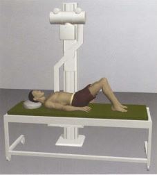
37105-4: XR Lumbar spine AP and Lateral and Spot Additionally, you can get information about the “37105-4” LOINC code in TXT format. This projection shows an orthogonal view of the AP/PA view and is utilized in many imaging contexts including trauma, postoperatively, and for chronic conditions. This view is also ideal in characterizing spinal alignment. Note: Ideally, spinal imaging should be taken erect in the non-trauma setting to give a functional overview of the lumbar spine. Lumbar Spine AP or PA. Purpose and Structures Shown A basic view of the lumbar spine. Position of patient Supine or prone. Injured patients should NOT be turned over. In trauma patients, a lumbar spine X-ray is done in the AP or PA position with minimal movement of the patient. Position of part The patient’s knees are bent to ensure the back. Start studying XR Positioning - Cervical Spine. Learn vocabulary, terms, and more with flashcards, games, and other study tools. AP, Lateral, R/L oblique, flexion, extension. Lateral Cervical structures. Pt stands in AP position- rotates 45 degree away from Bucky - rotate head so parallel w/ cassette- elevate chin-HOLD. The AP view will give the doctor a front-to-back picture of your back (where you facing the front of the x-ray machine) and lateral views give a side-to-side view (where you face sideways from the x-ray camera). In many cases, your doctor many order images that show weight-bearing, as this can provide a more accurate representation of your.
- XR ORDERING GUIDE - Providence Health & Services
- Xr L Spine Ap And Lateral
- Lateral C Spine Xr
- Xr Lumbar Spine Ap Lateral Flexion And Extension
- Thoracic Spine Ap And Lateral

XR ORDERING GUIDE - Providence Health & Services
In the context of trauma similar principles apply to imaging both the Thoracic spine (T-spine) and the Lumbar spine (L-spine). The plain X-ray anatomy and appearances of injuries to both these areas are discussed together.
Xr L Spine Ap And Lateral

Incorrect management of patients with spinal injury may cause or worsen neurological deficit. Therefore, patients with suspected spinal injury should be managed by experienced clinicians in accordance with local and national clinical guidelines. Imaging should not delay resuscitation.
Further imaging with CT or MRI (not discussed) is often appropriate in the context of a high risk injury, neurological deficit, limited clinical examination, or where there are unclear X-ray findings.
Good views of the T-spine and L-spine are difficult to achieve in the context of trauma. Clinical assessment is also often limited by distracting injuries or reduced consciousness. The clinico-radiological assessment of suspected T-spine or L-spine injuries therefore depends on careful consideration of both the clinical and radiological findings.
Thoracic spine - Standard views
Lateral C Spine Xr
AP and Lateral - Assess both views systematically (see box).
Xr Lumbar Spine Ap Lateral Flexion And Extension
Images of the thoracic and lumbar spine are often large and the bones should be scrutinised in detail (see images below).
Thoracic Spine Ap And Lateral
Note: The upper T-spine may not be visible on the lateral view - if injury is suspected here then a swimmer's view may be helpful - (see Cervical spine - Normal).